Home

Kocsma hegy Tenyésztés raman mapping Másodszor nyugalom vidék

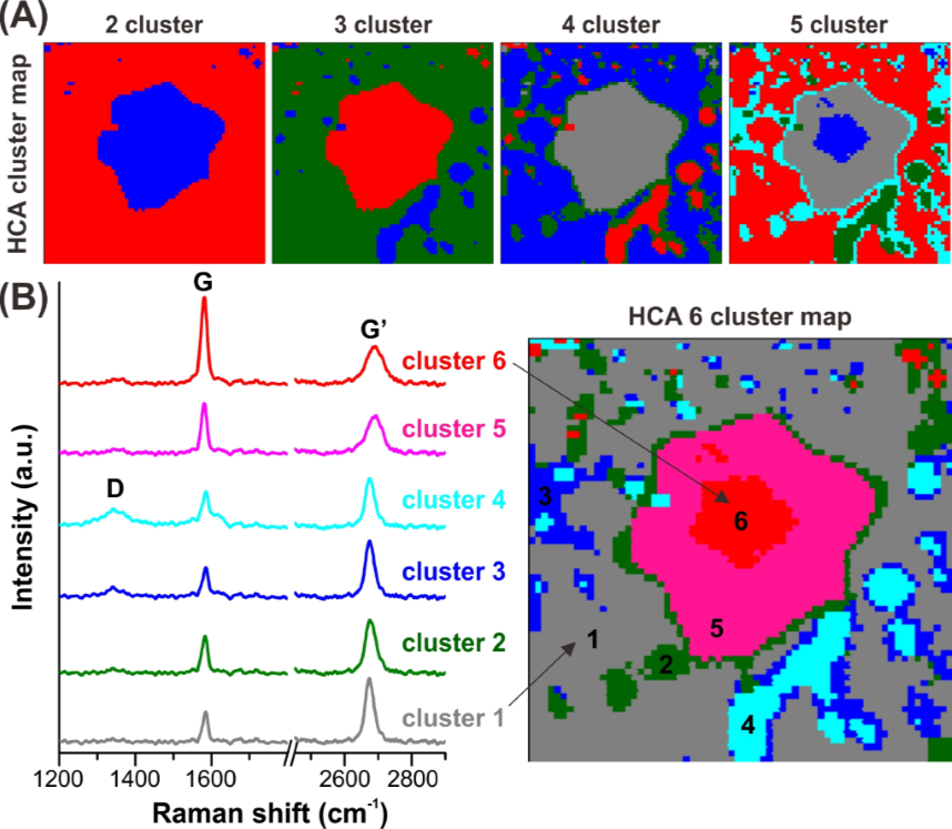
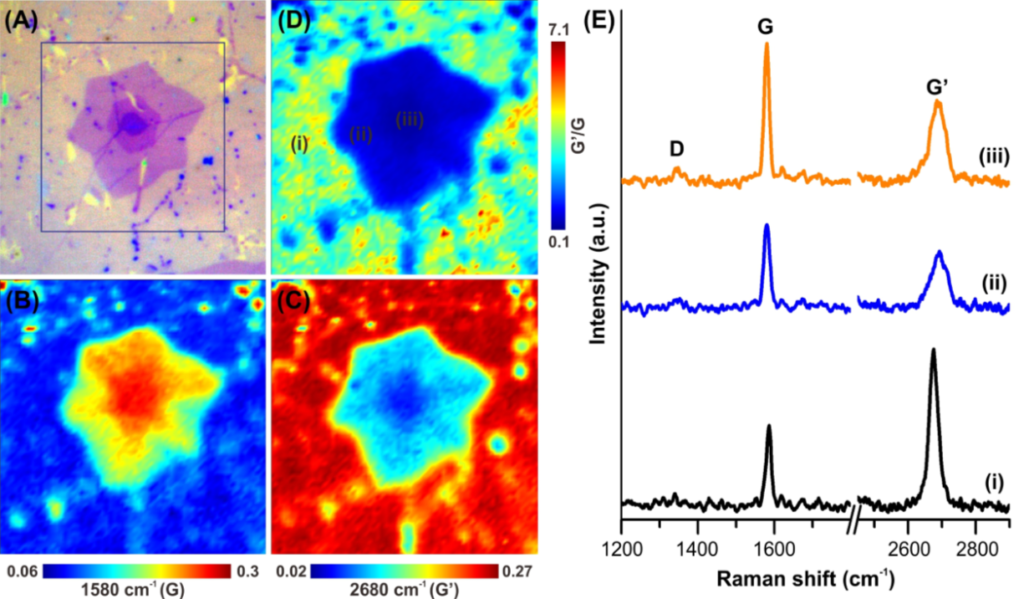
Spectroscopic Raman mapping of graphene grains and grain boundaries :... |  Download Scientific Diagram
Spectroscopic Raman mapping of graphene grains and grain boundaries :... | Download Scientific Diagram

Raman imaging through multimode sapphire fiber

Raman Mapping for the Investigation of Nano-phased Materials | SpringerLink

Raman mapping for single layer graphene - TechnoSpex Singapore

Raman imaging of a millimetre-sized core-shell SiO2@TiO2 sphere

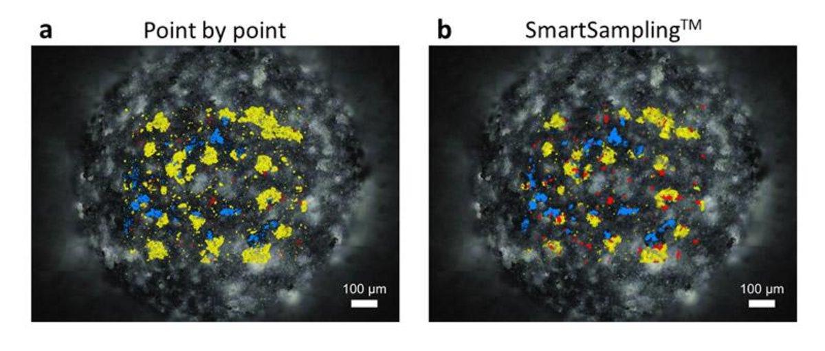
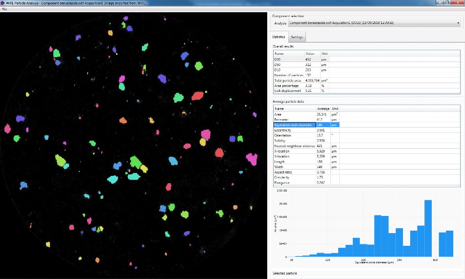
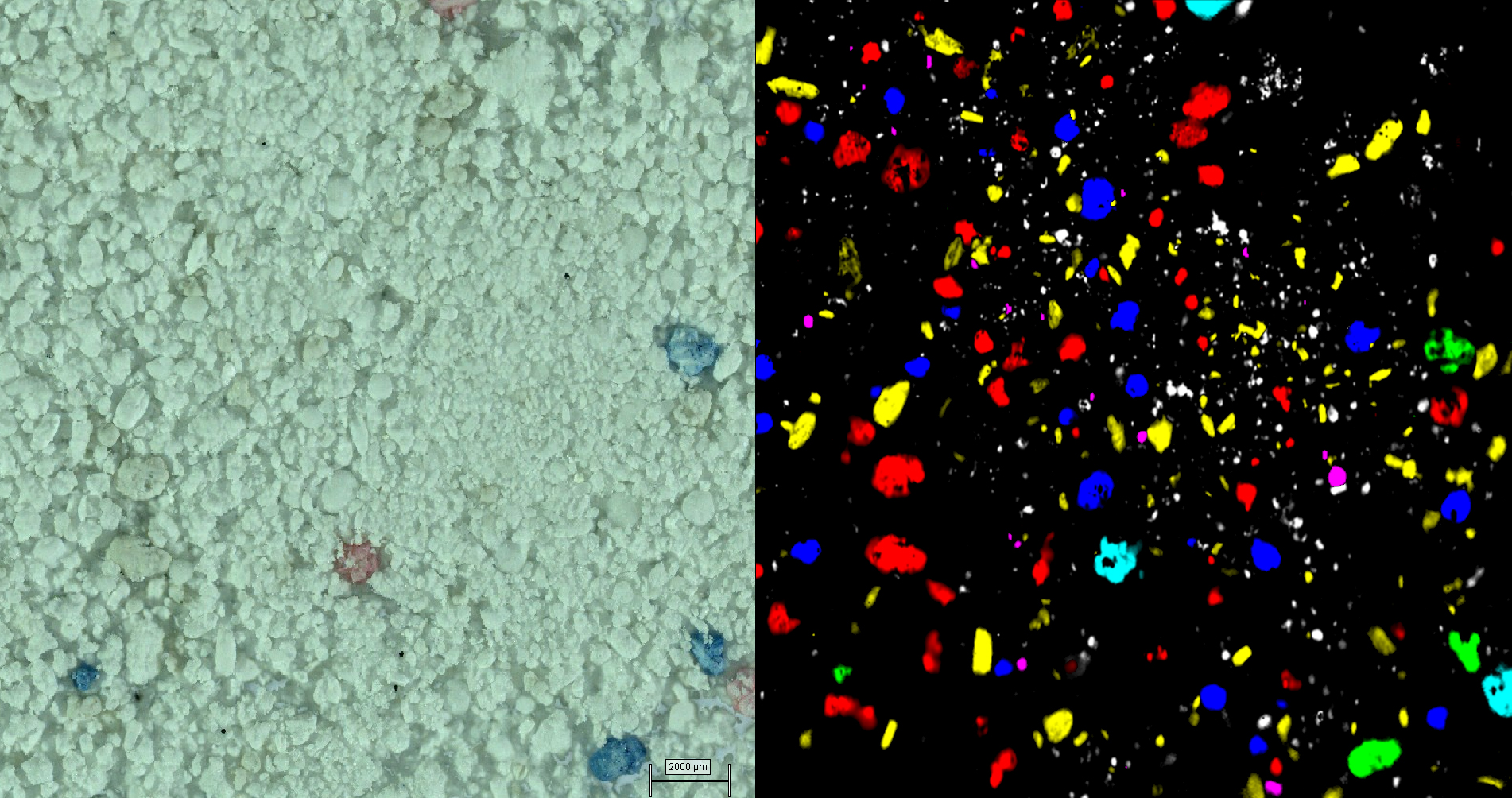
Using Raman Imaging for Deformulating Drugs

Petrographical and Mineralogical Applications of Raman Mapping | IntechOpen

Quantification and spatial distribution of salicylic acid in film tablets  using FT-Raman mapping with multivariate curve resolution - ScienceDirect
Quantification and spatial distribution of salicylic acid in film tablets using FT-Raman mapping with multivariate curve resolution - ScienceDirect

NT-MDT S.I. on Twitter: "Journal of Physical Chemistry C: Insights into the  interaction of Graphene Oxide and adsorbed RhB by Raman spectral  deconvoluted scanning. #Raman mapping done by SPECTRA II  https://t.co/x4P5C0rgzC . . #
NT-MDT S.I. on Twitter: "Journal of Physical Chemistry C: Insights into the interaction of Graphene Oxide and adsorbed RhB by Raman spectral deconvoluted scanning. #Raman mapping done by SPECTRA II https://t.co/x4P5C0rgzC . . #

Ultra-Fast Raman Mapping on Graphene - Live Demo - YouTube

Raman mapping images of phenylalanine activity: (a,b) with the 1005 cm... |  Download Scientific Diagram
Raman mapping images of phenylalanine activity: (a,b) with the 1005 cm... | Download Scientific Diagram

Raman Mapping of Lithiation Process on Graphene

Raman spectroscopy - Wikipedia

Raman mapping of the graphene film grown on the cemented carbide... |  Download Scientific Diagram
Raman mapping of the graphene film grown on the cemented carbide... | Download Scientific Diagram

Effect of graphene grain boundaries on MoS<sub>2</sub>/graphene  heterostructures
Effect of graphene grain boundaries on MoS<sub>2</sub>/graphene heterostructures

Raman images explained

Raman mapping investigation of chemical vapor deposition-fabricated twisted  bilayer graphene with irregular grains - Physical Chemistry Chemical  Physics (RSC Publishing)
Raman mapping investigation of chemical vapor deposition-fabricated twisted bilayer graphene with irregular grains - Physical Chemistry Chemical Physics (RSC Publishing)
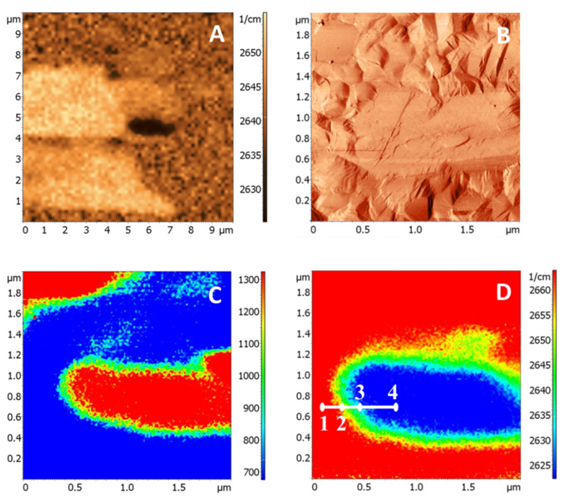
Tip-enhanced Raman mapping (TERM) of single-walled carbon nanotubes and  graphene | Spectroscopy Europe/World
Tip-enhanced Raman mapping (TERM) of single-walled carbon nanotubes and graphene | Spectroscopy Europe/World

Real time Raman imaging to understand dissolution performance of amorphous  solid dispersions - ScienceDirect
Real time Raman imaging to understand dissolution performance of amorphous solid dispersions - ScienceDirect

Raman mapping analysis of graphene film grown near the boundary of Cu... |  Download Scientific Diagram
Raman mapping analysis of graphene film grown near the boundary of Cu... | Download Scientific Diagram

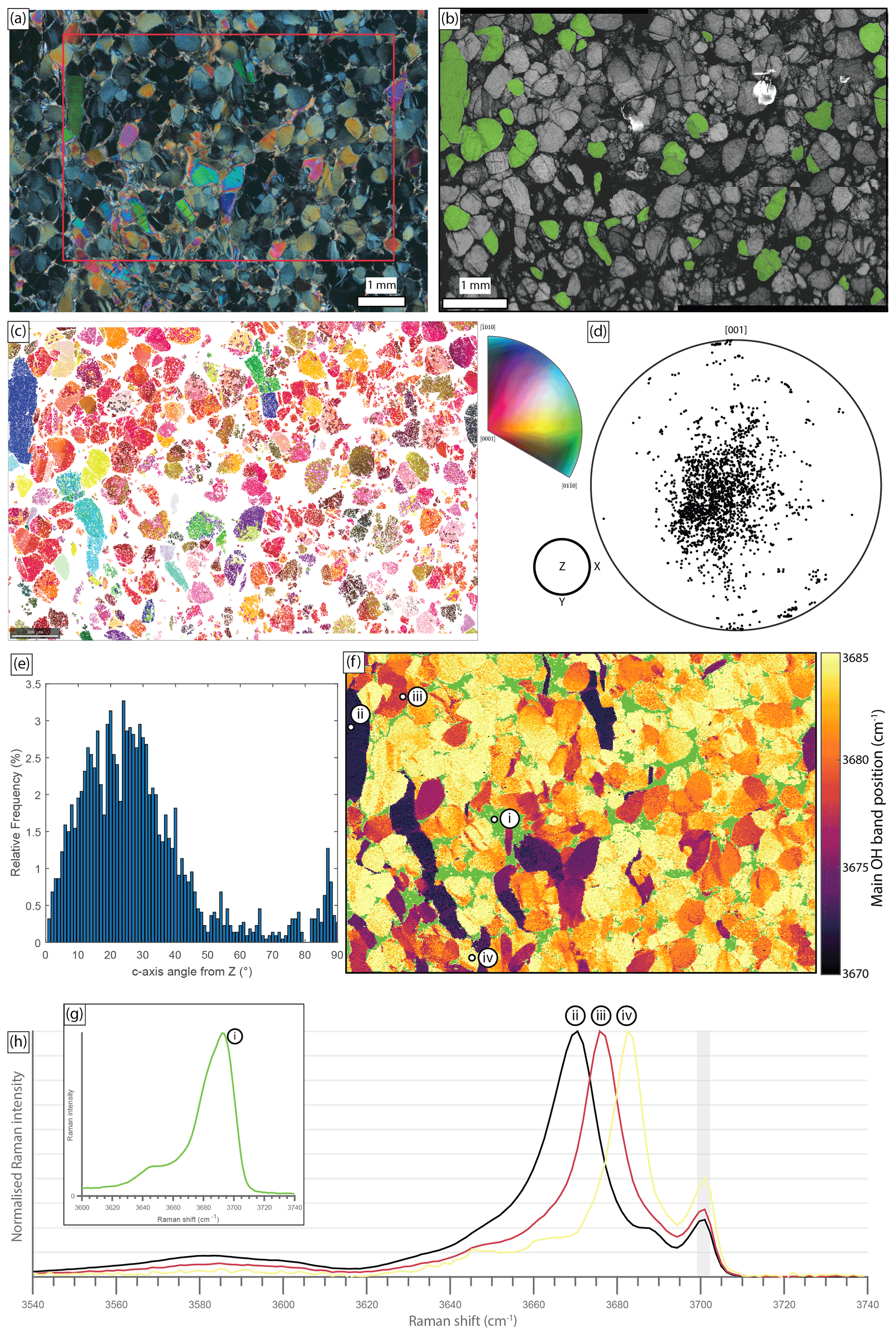
EJM - Crystallographic orientation mapping of lizardite serpentinite by  Raman spectroscopy
EJM - Crystallographic orientation mapping of lizardite serpentinite by Raman spectroscopy

Raman mapping for single layer graphene - TechnoSpex Singapore

Fast and quantitative 2D and 3D orientation mapping using Raman microscopy  | Nature Communications
Fast and quantitative 2D and 3D orientation mapping using Raman microscopy | Nature Communications